

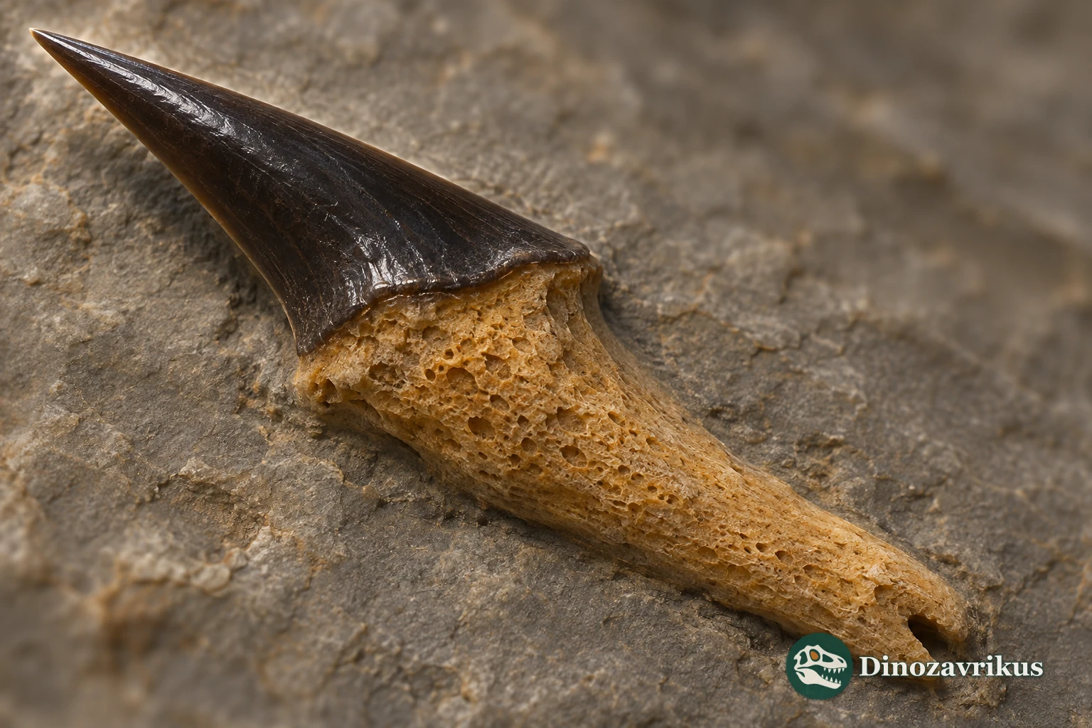
Dalpiazia was a Late Cretaceous sclerorhynchoid ray. The genus was named from a denticle that projected from the side of an elongated snout. The cleft in its base and the material inside it proved as informative as the outline of its cutting crown.
Quick facts
| Family | Sclerorhynchidae |
|---|---|
| Type species | Dalpiazia stromeri |
| Age of principal records | Maastrichtian |
| Type locality | Tripolitania, Libya |
| Fossils | Rostral denticles and oral teeth |
| Body length | Undetermined |
| Type collection | Sapienza University of Rome, MPUR ve.4465 |
What can the fossils tell us?
The Tripolitanian type preserves the crown and the part that attached to the snout. It offers no direct measurement of rostrum length.
Henri Cappetta recorded the notch and a small projection by the rear cutting edge on specimen MEN 9.
Enameloid reached about 250 micrometres along the edges of a denticle from Benguerir. The measurement applies to the sectioned specimen.
A separate genus
Giuseppe Checchia-Rispoli described Dalpiazia stromeri in 1933 from the upper Maastrichtian of Sofeggin in Tripolitania, Libya. The holotype, MPUR ve.4465 of the Sanfilippo collection, is held at the Palaeontological Museum of Sapienza University of Rome. Camille Arambourg subsequently grouped it with Onchosaurus on the strength of external similarities. In his 1972 study of the Iullemmeden Basin fish fauna, Henri Cappetta distinguished the two by the microstructure of their dentine and retained Dalpiazia.
Cappetta also treated material previously called Onchosaurus maroccanus and Onchosaurus manzadinensis as the same species. His account lists Maastrichtian occurrences in Libya, Morocco, Congo, Nigeria and Niger, linked by comparison of isolated denticles.
The Mentess denticle
In Cappetta’s most complete specimen from Mentess, Niger, the narrowing triangular cap has two nearly straight cutting edges. A small projection sits at the base of the shorter rear edge. The flattened attachment widens towards a deeply notched base with grooves along its margin. Broken specimens expose a wide cavity in the attachment.
The long, narrow crowns of the Moroccan sclerorhynchoid Ctenopristis sat on comparatively shallow bases. In Dalpiazia, the base extends farther and widens towards its attachment. That distinction survives in loose denticles even when the snout itself is missing.
Inside a denticle
In 2026 Endy Chateauneuf-Gautier and Guillaume Guinot examined denticles from bed C3 and oral teeth from bed C2 at Benguerir. In the transverse section of denticle BEG.C3-10, its hard enameloid coating measured 60–75 micrometres along the broad faces and about 250 micrometres at the cutting edges. Some crystallite bundles lie across the direction of the edges.
A longitudinal section of another denticle showed that the arrangement of the bundles also changed from the base to the tip. A sectioned oral tooth preserves a distinct vertical ridge on the outer side of its crown. These details describe individual elements. The full arrangement of the denticles and how the ray used its saw to catch prey remain unresolved.
Frequently asked questions
Where did the type specimen come from?
It came from upper Maastrichtian deposits in Tripolitania, in what is now western Libya.
Did the denticles form part of the jaws?
They lined the sides of the rostrum in front of the mouth. The fossil record also includes separate oral teeth.
Why has Dalpiazia been separated from Onchosaurus?
Cappetta found differences in the internal dentine structure despite similarities in the external shape.
Is an entire rostrum known?
The described fossils preserve individual denticles. Their total number and overall rostrum length are unresolved.