
Eoscapherpeton asiaticum was an aquatic salamander in the Late Cretaceous of Central Asia and an early member of the cryptobranchid lineage. Thousands of bones from Uzbekistan's Bissekty Formation offer an unusually rich record. They do not form one perfect skeleton: skull elements, vertebrae and limb bones from different individuals allow separate studies of anatomy, growth and injury.
Its place in the ancient amphibian catalogue reflects that evidence. Recent skull tomography also shows that its head differed from the flatter skulls of living giant salamanders.
Quick facts
| Scientific name | Eoscapherpeton asiaticum |
|---|---|
| Group | Early cryptobranchid salamander |
| Age | Turonian, Late Cretaceous, for the Bissekty sample |
| Key locality | Dzharakuduk, Bissekty Formation, Uzbekistan |
| Historical type | Left maxilla K77-1 in the original description |
| Sample | Thousands of isolated skeletal elements, not complete skeletons |
| Estimated length | Up to about 50–60 cm by comparison, not direct whole-skeleton measurement |
| Methods | Skull CT, femoral histology and palaeopathology |
What can the fossils tell us?
Several CT-scanned specimens show neurovascular canals and an ossified hyobranchial apparatus. Active aquatic predation and lateral-line prey detection are interpretations of those structures, not observed behaviour.
Different-sized femora show remodelling and changing vascularity. The 50–60 cm length is a comparative estimate, not a measurement of an articulated whole skeleton.
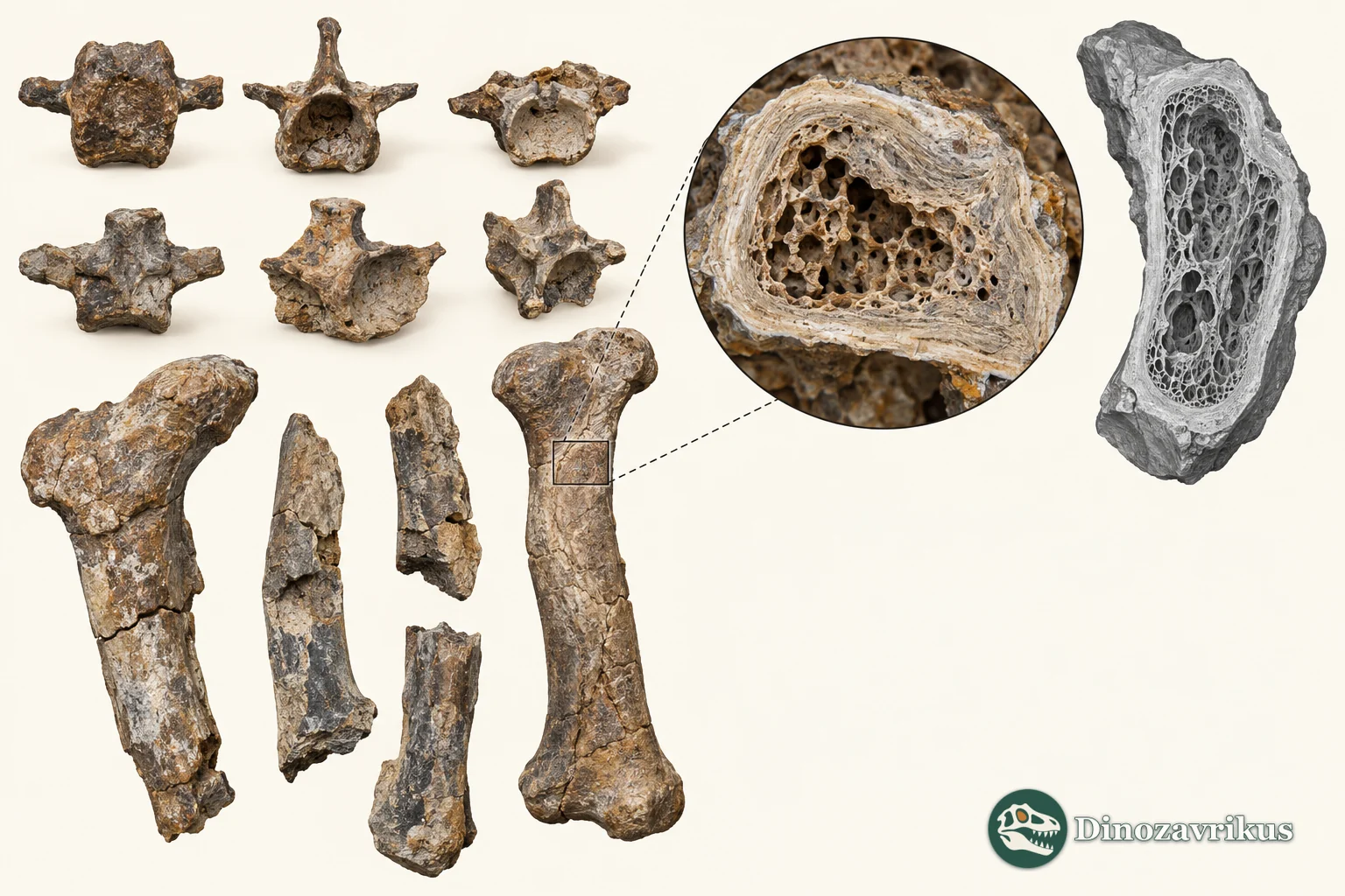
CT and sections distinguish jaw and femoral calluses from abnormal vertebral development. An individual injury's cause or a particular interaction between animals is not established.
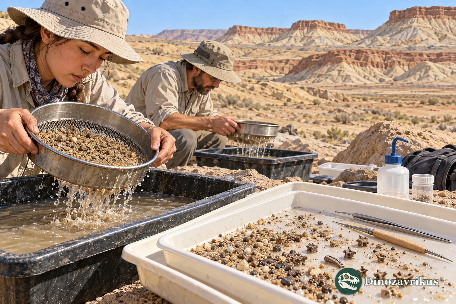
Expeditions recovered thousands of isolated Bissekty elements. The sample permits variation and pathology studies, but pieces cannot simply be joined into one animal or divided into an exact population count.
The Dzharakuduk sample and its limits
Lev Nesov's Central Asian fossil work established the name Eoscapherpeton asiaticum; the original type was recorded as left maxilla K77-1. Later collecting in the Kyzylkum Desert, including joint international expeditions from 1997 to 2006, recovered thousands of Bissekty bones. The well-studied Dzharakuduk material is Turonian in age. A heap of separated bones is not a count of complete skeletons or known individuals.
A pathology study selected 17 unusual jaw, vertebral and femoral elements held in the Zoological Institute collections in Saint Petersburg. Each specimen has a separate catalogue number. It would be wrong to treat the healed fracture in one femur as a trait of every salamander in the deposit.
A skull unlike a modern giant salamander's
Computed tomography of several cranial specimens revealed a skull less flattened from top to bottom than in living cryptobranchids. The authors documented an ossified hyobranchial apparatus and intricate neurovascular canals in the bones of the head, among other palatal and jaw characters. These structures support an interpretation of an active aquatic predator that could use a well-developed lateral-line system to find prey.
That is an inference from preserved structures and comparisons, not a record of a particular hunt. Nor should the powerful suction-feeding system of living giant salamanders be transferred wholesale to this earlier form: skull flattening and other features changed within the lineage.
Growing femora and an estimated body length
Researchers sectioned femora from different-sized animals. During growth, bone-tissue organisation, vascular networks and remodelling changed; calcified cartilage was progressively reduced in parts of the shaft. The series reveals skeletal development but does not supply a calibrated age in years for each individual.
The same work estimated body length up to roughly 50–60 cm by comparison. No one measured a complete articulated Eoscapherpeton skeleton of that exact length. The size of the particular animal in the cover remains a visual choice.
Healed fractures and developmental differences
Several femoral fragments and a jaw show healed breaks and callus. CT distinguishes the internal response from damage produced after burial. Some other bones suggest possible inflammation following trauma; the infectious agent itself was not found. A hemivertebra and several fused vertebrae were interpreted as developmental abnormalities.
Healing establishes that certain individuals survived long enough to lay down new bone. It does not identify what caused each injury. The authors discussed possible encounters among animals, but intraspecific aggression is one hypothesis rather than a witnessed event. Similarly, a few abnormal vertebrae in a large fossil sample should not be converted into an exact disease rate for the entire ancient population.
The Bissekty material belongs to the Cretaceous Period. A much older aquatic temnospondyl, Plagiosuchus, appears elsewhere in this catalogue; it was not a cryptobranchid salamander and never shared a waterway with Eoscapherpeton.
Frequently asked questions
Was Eoscapherpeton identical to living giant salamanders?
No. It belongs near the early cryptobranchid lineage, but its skull was less flattened. Modern species' feeding mechanics cannot simply be assigned to it.
Was a complete 60-centimetre skeleton found?
No. Up to about 50–60 cm is a comparative estimate based on separate bones from a large sample.
Can we tell why its bones broke?
Callus and CT show that some fractures healed. The actual event that caused each injury is unknown; conflict between individuals remains a possibility only.
What did skull CT add?
It revealed internal canals and hyobranchial details that are difficult to inspect from the surface. Researchers used those structures to infer aquatic prey detection, while keeping bone evidence distinct from behaviour.