

Roxochampsa paulistanus was a Late Cretaceous crocodylomorph from Brazil. It was first named from isolated material. Jaw fragments found elsewhere in São Paulo state carried the same distinctive pattern of ridges on the teeth and helped clarify its relationships.
Quick facts
| Species | Roxochampsa paulistanus |
|---|---|
| Group | Notosuchia, Itasuchidae |
| Age | Late Cretaceous |
| Region | Bauru Basin, São Paulo state, Brazil |
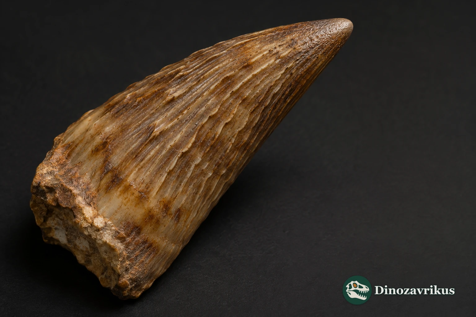
| Lectotype | DGM 259-R, part of a tooth crown |
| Jaw material | UFRJ-DG 451-R and UFRJ-DG 501-R |
What can the fossils tell us?
DGM 259-R was designated as the lectotype in 2018. The tibia from the original account came from a different site and cannot be securely identified with the teeth.
The scalloped edges are part of the enamel ornament. This is distinct from the true denticles found in some other crocodylomorphs.
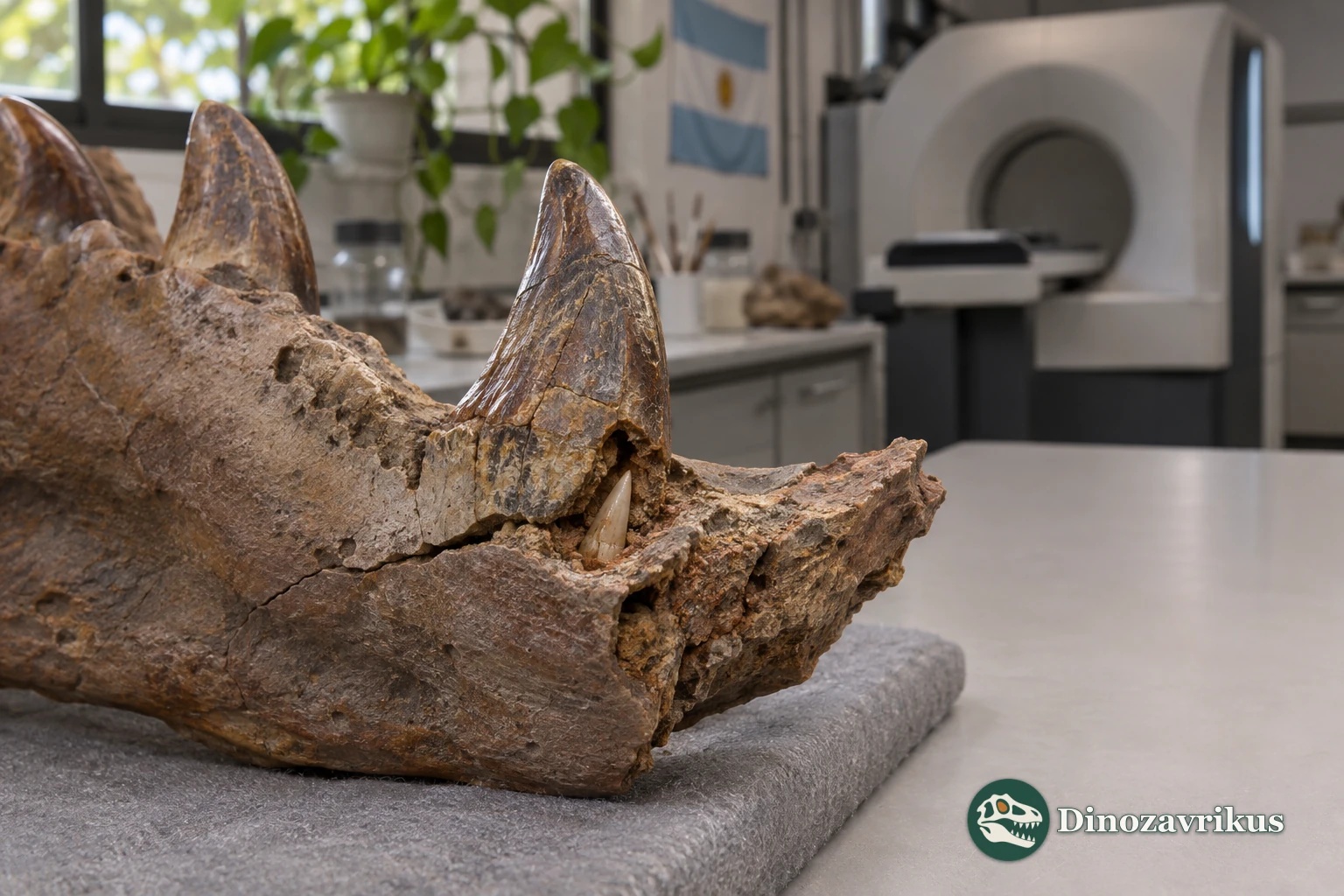
Micro-CT revealed unerupted teeth at the eleventh and thirteenth positions in UFRJ-DG 451-R. A single fossil cannot establish the rate of replacement.
Why the species received a new genus name
In 1936 Mathias de Oliveira Roxo named Goniopholis paulistanus from two teeth and a piece of a tibia. André Pinheiro and colleagues erected Roxochampsa in his honour in 2018. They selected the tooth DGM 259-R as the lectotype and DGM 258-R as the paralectotype. The tibia came from a separate locality and could not be identified to species.
The original teeth came from a railway cutting between Três Lagoas and Araçatuba and were assigned to the upper Adamantina Formation. The jaw fragments came from conglomerates of the Presidente Prudente Formation near Alfredo Marcondes. Their matching tooth ornament links the material.
Paired sockets and sculpted enamel
The jaws indicate at least nineteen dentary tooth positions per side. Sockets six and seven form a close pair, as do sockets eight and nine. Itasuchus also has closely paired positions. In Roxochampsa the final two teeth shared a groove, a feature that helps distinguish it from its relatives.
Raised, finely scalloped ridges run along the conical crowns. Some bear blunt overlapping hooks near the tip. The detail of those ridges, visible on both loose teeth and teeth in the jaws, was central to the 2018 reidentification.
Inside the fossil jaw
Micro-CT scans of UFRJ-DG 451-R show three complete sockets and replacement teeth at the eleventh and thirteenth positions. The teeth had formed beneath the working row, where they were obscured from ordinary view.
The rounder rear crowns have prompted suggestions that the animal could handle harder food. Specific prey and total body length remain uncertain because the material is mainly dental and mandibular.
Frequently asked questions
What does the lectotype preserve?
The pointed part of an isolated tooth crown, DGM 259-R.
Why was the tibia left unassigned?
It came from a different site and lacks evidence tying it to the type teeth.
How were the replacement teeth detected?
Micro-CT revealed them beneath the working row at the eleventh and thirteenth tooth positions.
What is different about the last two teeth?
They occupied a common groove in the lower jaw, rather than two completely separate sockets.