

A fossil may hide anatomy that cannot be seen on its surface or in a single radiograph. Synchrotron tomography records many X-ray projections around a specimen and reconstructs them as a digital volume. Researchers can inspect virtual slices, separate fossil from matrix where the signals allow it, and measure structures without cutting through a rare specimen.
A synchrotron does not simply “photograph through a fossil” in one exposure. It is an X-ray source associated with a large accelerator facility; tomography and digital reconstruction are separate parts of the experiment. As with other ways of studying fossils, the useful result depends on the material and the question.
Where synchrotron X-rays come from
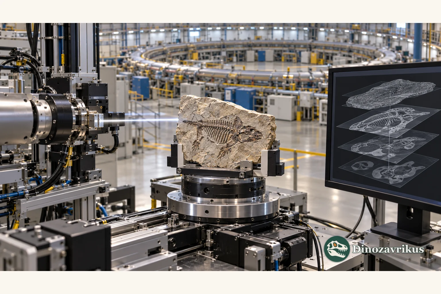
In a storage ring, charged particles travel at very high speed. Magnets bend their paths and produce intense radiation. At a dedicated beamline, X-rays are directed through a mounted specimen and recorded by a detector. For tomography, the specimen is rotated and imaged from many angles. A reconstruction algorithm turns those projections into a stack of two-dimensional slices that can also be combined into a three-dimensional model.
The brightness of the beam can support a large number of projections with a strong signal, while the beam’s coherence can enable contrast methods unavailable on many laboratory scanners. Brightness is not the same thing as a guarantee of anatomical clarity: the fossil and its surrounding rock must produce distinguishable signals, and the detector and geometry must suit the sample.
Why phase contrast can reveal a boundary
Conventional X-ray images are based mainly on how much a material attenuates the beam. If fossil and matrix have similar composition or density, their signals may overlap. X-ray waves also change phase as they pass through material. With a coherent beam, small phase shifts can create visible edge effects at boundaries between materials.
Propagation-based phase contrast can help where absorption contrast is weak but interfaces remain detectable. A methodological comparison tested laboratory and synchrotron setups on the fossil Corumbella werneri and showed that imaging configurations differ in the internal structures they reveal. It is an example of matching a setup to a specimen, not evidence that a synchrotron always outperforms every other scanner.
What a digital volume can answer
Internal imaging can reveal the arrangement of small skeletal elements, cavities and canals, tooth tissues, or structures enclosed within an embryo. In a study of fossil embryos from the late Neoproterozoic and earliest Cambrian, synchrotron tomography produced three-dimensional records at submicrometre scale and helped distinguish phosphate phases involved in preservation. Those results depend on the preservation of those particular fossils; they cannot be assumed for every specimen.
Virtual slices also let researchers revisit the same object with different segmentation choices. If data and methods are shared, other teams can inspect boundaries, compare alternative models and measure features that are difficult to assess from the exterior. The 3D rendering is still a processed representation of measurements, not independent proof of every anatomical interpretation.
How it differs from laboratory micro-CT
Laboratory micro-CT generates X-rays inside the scanner and is available in many museum and university labs. It is often suitable for bones, teeth and small blocks. Synchrotron beamlines are concentrated in large research facilities, where access is scheduled among many fields. Their advantages matter when a study needs high intensity, rapid acquisition, a particular energy range or phase contrast.
The choice is not a simple “synchrotron is better” rule. Resolution depends on sample size, detector, geometry, exposure and processing. A small fragment may be scanned in great detail, while a large block requires a compromise. If fossil and matrix produce nearly identical signals, a powerful source still may not give a clean boundary. For other small remains, screening sediment for microfossils may answer a different question more effectively.
Limits and interpretation
A scan cannot restore tissues that did not survive, and it cannot identify a species merely because a reconstruction looks vivid. Cracks, mineral replacement, burial deformation and an uneven matrix can imitate or obscure anatomical boundaries. Segmentation is performed by researchers or algorithms using selected parameters; ambiguous contrast may lead to different plausible models.
Although tomography is commonly treated as non-destructive, rare collection objects are assessed before an experiment. Size, mass, mounting, exposure and storage requirements all matter. Sometimes only a fragment is scanned because a whole specimen is too large or cannot fit within the field of view.
A useful report states which specimen was scanned, the imaging mode and resolution, and how the volume was reconstructed. Readers should distinguish directly visible structure from interpretation and from colours chosen for display. Synchrotron imaging adds a powerful way to observe fossils, but conclusions still need to be checked against anatomy, geology and other evidence.
Frequently asked questions
Does synchrotron scanning damage a fossil?
Tomography is generally used as a non-destructive method: researchers do not have to cut the specimen to obtain internal slices. The specific material, mounting and exposure still need to be assessed; no single guarantee applies to every museum object.
Is synchrotron tomography always better than laboratory micro-CT?
No. A synchrotron is useful when a project needs particular intensity, contrast, speed or phase imaging, while laboratory micro-CT is more accessible and often sufficient. The specimen and research question determine the choice.
Can a scan reveal soft tissue in every fossil?
No. Imaging can show only structures that survived and produce a distinguishable signal. It cannot recreate tissue that has been lost or resolve a boundary when fossil and matrix are indistinguishable.
What does phase contrast mean in simple terms?
An X-ray wave changes as it passes through different materials. Phase-contrast imaging uses some of those changes to make boundaries more visible, even when the materials absorb similar amounts of X-rays.